There are four laboratory space in Yap research lab:
- The main laboratory (B031) houses most of the major materials synthesis facilities.
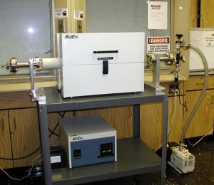
- The chemistry laboratory (B012) is equipped with a 1500 °C chemical vapor deposition system and wet batch facility.
- The spectroscopy laboratory (B034) features all the major characterization systems, including a micro-Raman spectrometer, a micro-FTIR spectrometer, a micro-probe station, a solar simulator, a UV-VIS spectrometer, etc.
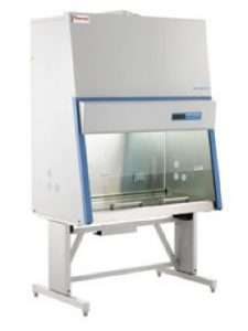
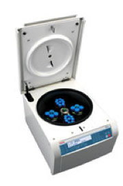
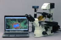
- The biochemical physics (ChemBioPhys) laboratory (B009) is specifically designed for chemical, biological, and electrochemical experiments. It has a controlled-atmosphere glove box, a computer-controlled electrochemical workstation, and a comprehensive molecular biology facility with a Class A2 Biological Safety Cabinet, a CO2 Incubator, a Refrigerated Centrifuge, a Freezer (-30 °C), a Phase-contrast Fluorescence Inverted Microscope with digital camera, a Sterilizer, a Cryogenic Storage system, etc.
- Yap is the investigator of NSF Major Research Instrumentation (MRI) grants for an In-Situ AFM/STM-TEM System (2008), and a High Resolution Transmission Electron Microscope for In Situ Microscopy (2014).
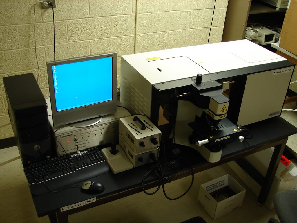
Jobin-Yvon LabRAM HR800 Raman Spectrometer
This Raman spectrometer can be operated with one of the three excitation laser lines: HeNe laser (wavelength = 633 nm), and NeCd laser (wavelengths = 442 nm and 325 nm). This is a high-resolution system (~ 0.1 cm-1) with edge filters that enable the detection of Stokes Raman scattering under a confocal optical microscope. These high-quality filter enable excellent laser line cut-off (~80cm-1 from all laser lines), which is desirable for the detection of interesting Raman scattering including Radial Breathing Mode (RBM) of single wall carbon nanotubes (SWCNTs). This microscope has a high-precision x-y mapping stage with minimum steping of 100 nm. This sample stage in conjugate with a computer aided CCD camera allowed three dimensional (3D) sample mapping. This Raman system is also configurated for photoluminescence (PL) experiments with the UV HeCd excitation line. The detection range of the CCD sensor is between ~380–900 nm. In addition, this system is equiped with the Linkam cooling and heating stages and will enable Raman scattering to be characterized at desired ambient and temperatures between –196°C to 1500 °C.
USE RATE: This spectrometer is now opened for on-campus and off-campus users. The present rate is $80/hour without an assistant. Additional labor cost will be charged to user’s index when assistance is needed. Please contact Professor Yoke Khin Yap (E-mail: ykyap@mtu.edu) for details.
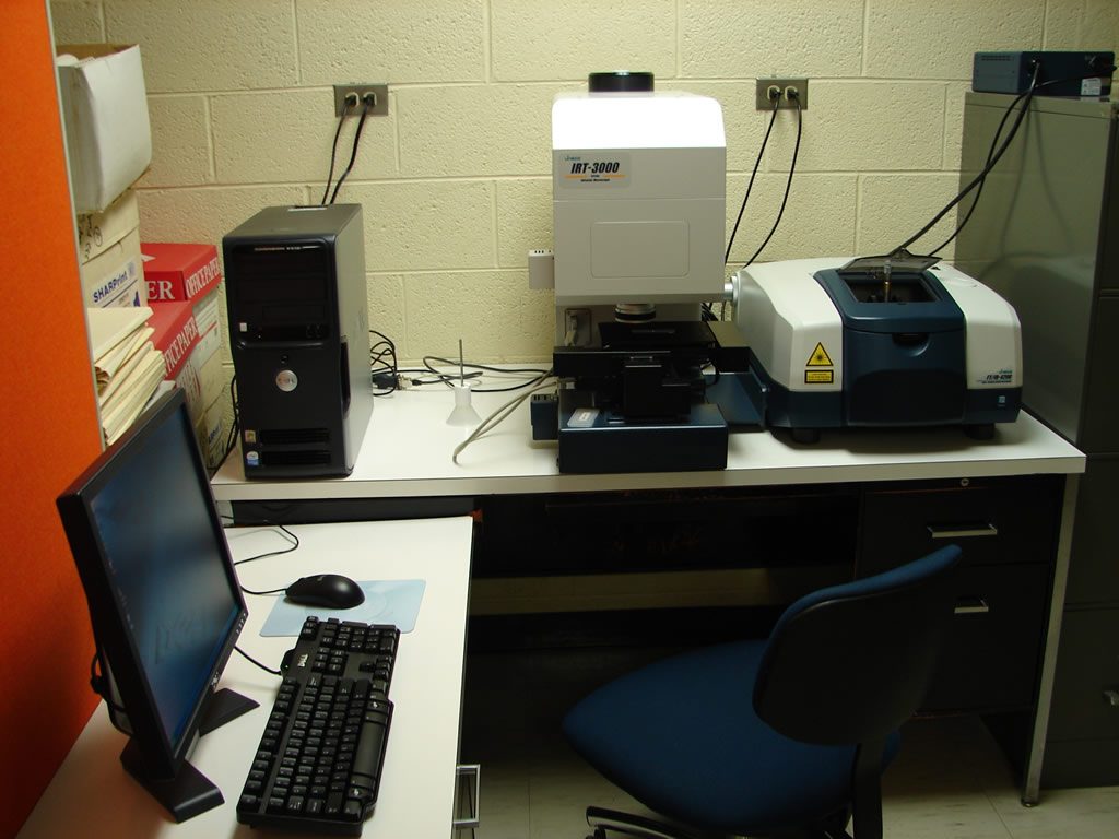
Jasco Fourier Transformed Infrared (FTIR) Spectrometer
Last updated July 23, 2017
This FTIR system consists of a FT/IR-4200 spectrometer and an IRT-3000 FTIR microscope. IR spectra can be acquired macroscopically in the FT/IR-4200 spectrometer or microscopically under the microscope. The spectral range of the spectrometer is 7800 – 350 cm-1 with step selectable resolutions of 16, 8, 4, 2, 1 and 0.5 cm-1. A minimum signal- to-noise ratio of 30,000:1, RMS 120,000:1 can be expected. This spectrometer has a 90-degree Michelson interferometer utilizing retro-reflecting corner cube mirrors and a ceramic mid-infrared source with a 2-stage power supply. The microscope equipped with an automated X-Y-Z microscope stage with sample autofocus capability. It is connected to a mid band MCT detector with a spectral range of 5000 – 550 cm-1. Its sample aperture is computer controlled and capable of infrared spectral measurements with a spatial resolution of 10 microns. This microscope is interfaced with a mapping software includes the ability to collect line and area maps. Spectral mapping data is processed and available as 3-D, contour, line and intensity mapping images.
USE RATE: This spectrometer is opened for on-campus and off-campus users under a collaboration basis. No charge will be applied. Please contact Professor Yoke Khin Yap (E-mail: ykyap@mtu.edu) for details.
Cascade Microtech M150 Probe Station
 This is a benchtop probing system for electrical measurement of microelectronic devices with wafer size up to 150 mm in diameter. This probe station has a fixed platen and features a Vacuum Glide stage. This stage provides non leadscrew-based free-form gross positioning of the wafer chuck. This system also has a video-ready, stereozoom microscope kit (Leica S6D) that allow device monitoring from the ccd camera-monitor system.
The Biochemical Physics (BioChemPhys) Facility
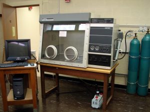
This laboratory is equip with a controlled atmosphere glove box (Labconco Protector), and an electrochemical workstation (CHI 660B). The glove box has a regenerative drying train (AtmosPure) that help to maintain the glove box atmosphere of < 5 ppm water and <1 ppm oxygen. The workstation is computer controlled with a Windows-based electrochemical instrument for performing measurements including various voltammetry, amperometry, chronopotentiometry, and potentiometry, etc.
This lab space also equipped with a comprehensive cell culture facility including a class II biological safety cabinet, a CO2 Incubator, Refrigerated Centrifuge, Fluorescence Inverted Microscope with digital camera, etc.
The Materials Synthesis Facility
Pulsed-Laser Deposition (PLD) system
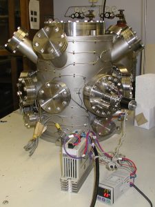
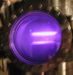
Plasma-enhanced Chemical Vapor Deposition (CVD) system
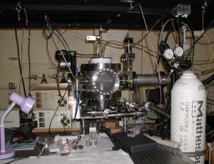
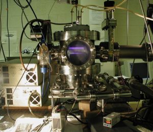
Thermal Chemical Vapor Deposition (CVD) system
High Resolution Transmission Electron Microscope for In Situ Microscopy